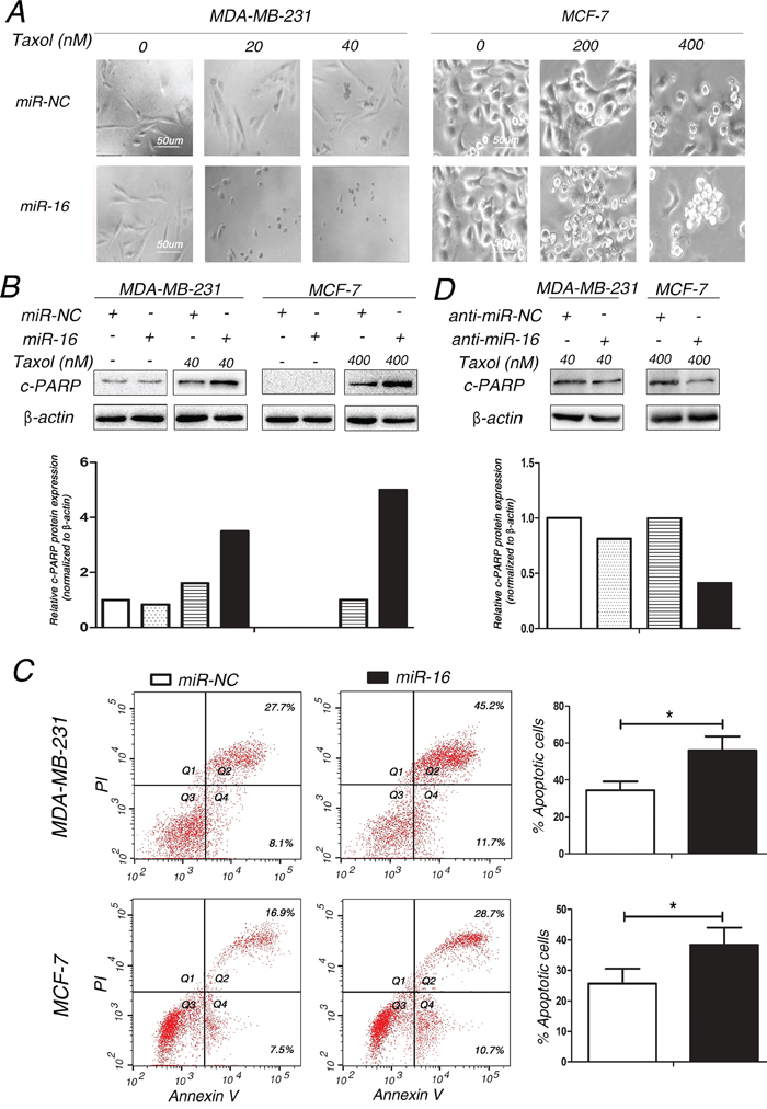
Figure 2. Involvement of miR-16 in Taxol-induced apoptosis in breast cancer cells.

A. MDA-MB-231 and MCF-7 cells transfected with 50 nM miR-16 mimics or 50 nM miR-NC were treated with 0, 20, 40 nM (MDA-MB-231) or 0, 200, 400 nM (MCF-7) Taxol for 48 h. The cellular morphologies were visualized using a phase-contrast microscope. B-C. MDA-MB-231 and MCF-7 cells were transfected with 50 nM miR-NC or miR-16 mimics and then treated with 40 and 400 nM Taxol for 48 h, respectively. Cell lysates were extracted for western blotting using an antibody against c-PARP (B), or cells were collected for annexin V staining and flow cytometry assays (C). The gray density was quantified using the ImageJ software and normalized to β-actin. The percentage of apoptotic cells is represented in a bar diagram from three independent experiments. D. MDA-MB-231 and MCF-7 cells transfected with 100 nM anti-miR-NC or miR-16 inhibitor were treated with 40 and 400 nM Taxol for 48 h, respectively. Cell lysates were extracted for western blotting using an antibody against c-PARP. β-actin was used as an internal control. Columns, means of three independent experiments; bars, S.E. *, p<0.05, **, p<0.01.
