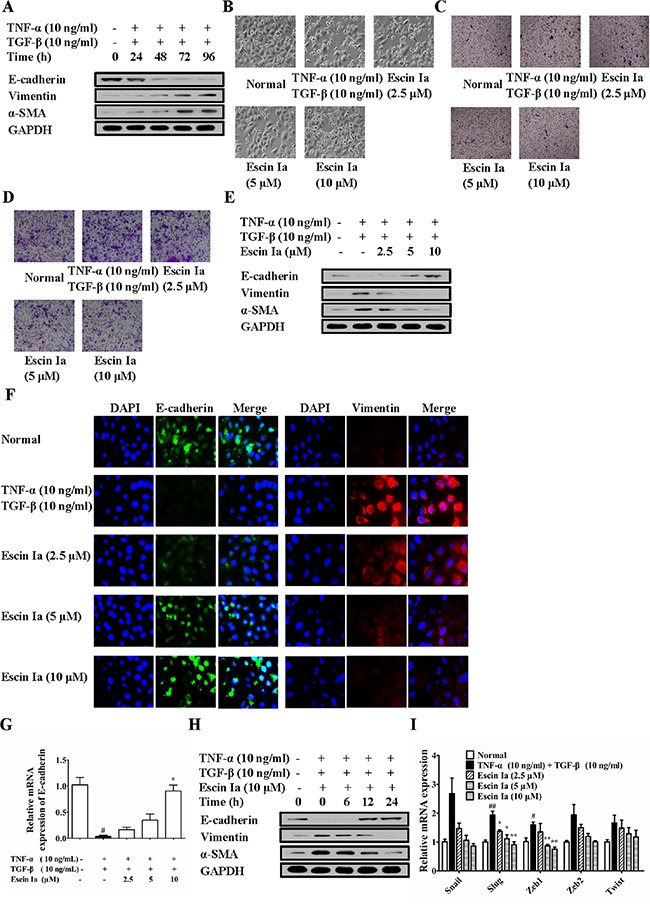
Figure 5. Effect of escin Ia on epithelial-mesenchymal transition in TNF-α/TGF-β-stimulated MCF-7 cells.

(A) MCF-7 cells were treated with TNF-α+TGF-β (10 ng/mL of each) for 0 h, 24 h, 48 h, 72 h, 96 h, and the total protein lysates were immunoblotted for E-cadherin, vimentin and α-SMA expressions. B-I, MCF-7 cells were incubated with TNF-α+TGF-β (10 ng/mL of each) plus escin Ia (2.5, 5, 10 μM) or escin Ia (10 μM) for the indicated internals. Cell morphology was recorded under an inverted microscope (magnification 200 ×) (B). Cell invasion was detected by using cell invasion assays (C). Cell migration was detected by using cell migration assay (D). The protein expressions of E-cadherin, vimentin and α-SMA were detected by using western blot analysis (E). The protein expressions of E-cadherin and vimentin were detected by using immunofluorescence assay (F). The mRNA expressions of E-cadherin, Snail, Slug, Zeb1, Zeb2, Twist were detected by using Q-PCR assay (G, I). Gene expressions were normalized to GAPDH. Time course of escin Ia on protein expressions of E-cadherin, vimentin and α-SMA were detected by using western blot analysis (H). The data were expressed as the means ± S.E.M. of three independent experiments. #p < 0.05, ##p < 0.01 vs. normal; *p < 0.05, **p < 0.01 vs. TNF-α (10 ng/mL) + TGF-β (10 ng/mL).
