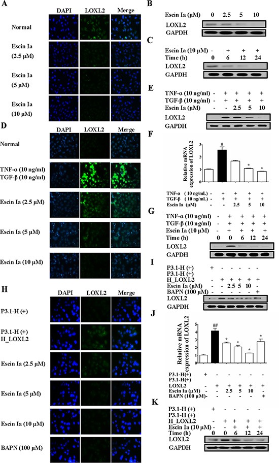
Figure 6. Effect of escin Ia on LOXL2 expression in MDA-MB-231 cells and TNF-α/TGF-β-stimulated or LOXL2-transfected MCF-7 cells.

(A–C) Effect of escin Ia on LOXL2 expression in MDA-MB-231 cells. MDA-MB-231 cells were treated with escin Ia (2.5, 5, 10 μM) or escin Ia (10 μM) for the indicated intervals. The protein expression of LOXL2 was detected by using immunofluorescence and western blot analysis (A, B). Time course of escin Ia on protein expression of LOXL2 was detected using western blot analysis (C); D-G, MCF-7 cells were incubated with TNF-α+TGF-β (10 ng/mL of each) plus escin Ia (2.5, 5, 10 μM) or escin Ia (10 μM) for the indicated internals. The protein and mRNA expression of LOXL2 was detected by using immunofluorescence, western blot and Q-PCR assays (D–F). Time course of escin Ia on protein expression of LOXL2 was detected using western blot analysis (G); H-K, MCF-7 cells were transiently transfected with a plasmid of LOXL2 (P3.1-H (+) H_LOXL2) plus escin Ia (2.5, 5, 10 μM), BAPN (100 μM) or escin Ia (10 μM) for the indicated internals. The protein expression of LOXL2 was detected by using immunofluorescence assays, western blot analysis and Q-PCR assays (H–J). Time course of escin Ia on protein expression of LOXL2 was detected by using western blot analysis (K). The data were expressed as the means ± S.E.M. of three independent experiments. #p < 0.05 vs. TNF-α (10 ng/mL) + TGF-β (10 ng/mL), ##p < 0.01 vs. p3.1-H (+); *p < 0.05 vs. TNF-α (10 ng/mL)+TGF-β (10 ng/mL), *p < 0.05 vs. p3.1-H(+) H_LOXL2.
