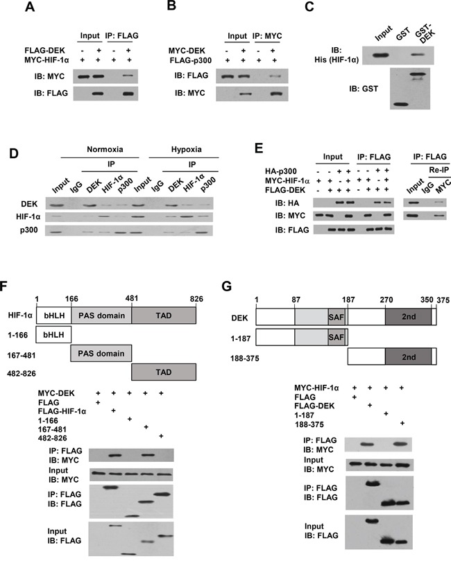
Figure 6. DEK, HIF-1α and p300 form a complex in vivo.

A. Co-IP assays with HEK293T cells cotransfected with MYC-tagged HIF-1α and FLAG-tagged DEK. Cell lysates were immunoprecipitated (IP) with anti-FLAG, followed by immunoblotting (IB) with the indicated antibodies. B. Co-IP assays with HEK293T cells cotransfected with MYC-tagged DEK and FLAG-tagged p300. Cell lysates were immunoprecipitated with anti-MYC, followed by immunoblotting with the indicated antibodies. C. GST pull-down analysis of direct interaction between DEK and HIF-1α. Purified His-tagged HIF-1α was incubated with purified GST-DEK or GST beads. Immunoblot with anti-His was performed. D. Reciprocal co-IP analysis of endogenous interactions among DEK, HIF-1α and p300 under normoxic or hypoxic conditions for 24 h. Immunoprecipitation were performed with antibodies against DEK, HIF-1α or p300, followed by immunoblot with the indicated antibodies. E. HEK293T cells were cotransfected with MYC-tagged HIF-1α, FLAG-tagged DEK and HA-tagged p300. Cell lysates were immunoprecipitated with anti-FLAG (1st IP). The immune complexes were eluted with FLAG peptide and re-immunoprecipitated (Re-IP) with anti-Myc or normal IgG, followed by immunoblotting with the indicated antibodies. F. HEK293T cells were cotransfected with MYC-tagged DEK and FLAG-tagged HIF-1α or its deletion mutants. Cell lysates were immunoprecipitated with anti-FLAG, followed by immunoblotting with the indicated antibodies. Schematic diagram of HIF-1α and its deletion mutants is shown. bHLH, basic-Helix-Loop-Helix; PAS, Per-ARNT-Sim; TAD, transactivation domain. G. HEK293T cells were cotransfected with MYC-tagged HIF-1α and FLAG-tagged DEK or its deletion mutants. Cell lysates were immunoprecipitated and analyzed as in A. Schematic diagram of DEK and its deletion mutants is shown. SAF, scaffold attachment factor (first DNA-binding domain); 2nd, second DNA-binding domain.
