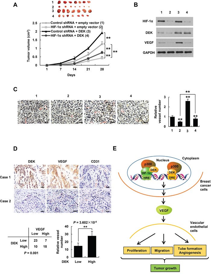
Figure 7. DEK controls tumor angiogenesis in HIF-1α-dependent and -independent manners.

A. Volume of xenograft tumors derived from MDA-MB-231 cells stably infected with lentiviruses carrying the indicated constructs. Data are shown as mean ± SD (n = 7). **P < 0.01 on day 28. B, C. Representative tumor tissues from A were subjected to immunoblot with the indicated antibodies (B) and immunohistochemical staining with an anti-CD31 antibody (C). Pictures from eight areas in each group were taken and the vessel number was manually counted (C). Scale bar: 50 μm. D. Representative immunohistochemical staining of DEK, VEGF and CD31 in human breast cancer samples. Scale bar: 25 μm. For the quantification of microvessel density, pictures from eight areas in each tissue were taken and the vessel number was counted. The correlation of DEK with VEGF or microvessel number (positive CD31 staining) is shown. The P value was generated using Pearson's χ2 test (DEK and VEGF) and Wilcoxon ranked sum test (DEK and CD31). E. Proposed model for DEK modulation of VEGF expression and tumor angiogenesis. DEK directly binds to the DRE of the VEGF promoter and indirectly binds to the HRE via its interaction with HIF-1α, and recruits the histone acetyltransferase p300 to both sites, resulting in increased VEGF transcription and secretion in breast cancer cells. The cancer cell-secreted VEGF promotes vascular endothelial cell proliferation, migration and tube formation as well as tumor angiogenesis and growth.
