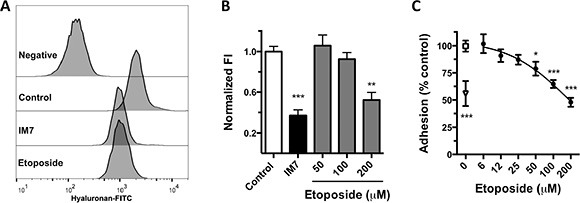
Figure 3. Inhibition of HA-CD44 binding by etoposide.

(A) Flow cytometry histograms of HA-FITC binding to MDA-MB-231 control cells (0.2% DMSO) or cells treated with anti-CD44 (mAb IM7) or 200 μM etoposide. Negative fluorescence consists of cells incubated with nonfluorescent HA. (B) Quantification of normalized fluorescence index (FI; see “Methods”) from 5 independent experiments (means ± SEM). **P < 0.01, ***P < 0.001 by Bonferroni's multiple comparisons test. (C) Cell adhesion of MDA-MB-231 cells to HA-coated microplates. Cells were treated with 0.2% DMSO (□), various concentrations of etoposide (·), or IM7 antibody (Δ). Data are means ± SEM from 3 independent experiments. *P < 0.05, ***P < 0.001 by Bonferroni's multiple comparisons test.
