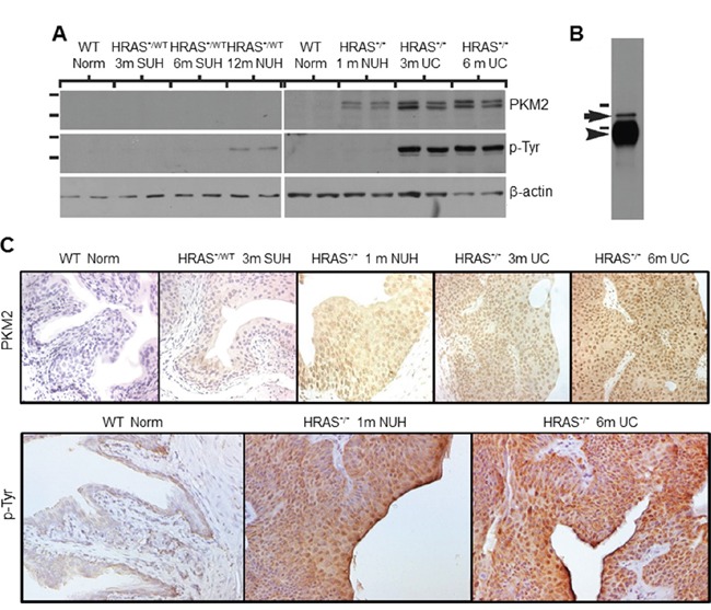
Figure 2. Expression levels and subcellular localization of PKM2 in low-grade urothelial lesions induced by activated HRAS.

A. Western blotting of total protein extracts of normal urothelial cells from wild-type mice (6 months of age), simple urothelial hyperplasia (SUH) from 3- and 6-month-old heterozygous Upk2-HRAS*/WT transgenic mice (abbreviated as HRAS*/WT), nodular urothelial hyperplasia (NUH) from 12-month-old heterozygous Upk2-HRAS*/WT transgenic mice and 1-month-old homozygous Upk2-HRAS*/* transgenic mice, and low-grade papillary urothelial carcinoma (UC) from 3- and 6-month-old homozygous Upk2-HRAS*/* transgenic mice, using anti-PKM2, anti-phosphotyrosine and anti-actin antibodies. Note the first appearance of PKM2 and phosphorylated PKM2 in nodular urothelial hyperplasia followed by an increased intensity in low-grade UC. The doublet of PKM2 most likely represented the non-phosphorylated and phosphorylated versions of PKM2. B. Immunoprecipitation (IP) using as starting materials total proteins from low-grade papillary UC (as shown in (A), last two lanes), using anti-phosphotyrosine antibody followed by Western blotting using anti-PKM2 antibody. Note the specific identification of the 60-kDa PKM2 (arrow). Arrowhead denotes the IgG heavy chain present in the IP product reactive with the secondary antibody during Western blotting. Short horizontal bars denote two of the molecular weight standards (top, 72-kDa; bottom, 55-kDa). C. Immunohistochemistry using anti-PKM2 and anti-phosphotyrosine showed that the expression of PKM2 and p-PKM2 corresponded well with that of the Western blotting and that prominent nuclear staining was present. Magnification of panels in C: 200x.
