
Figure 2. PAI-1 secreted from radioresistant cells under irradiation is a key paracrine factor in survival of radiosensitive cells in NSCLC.
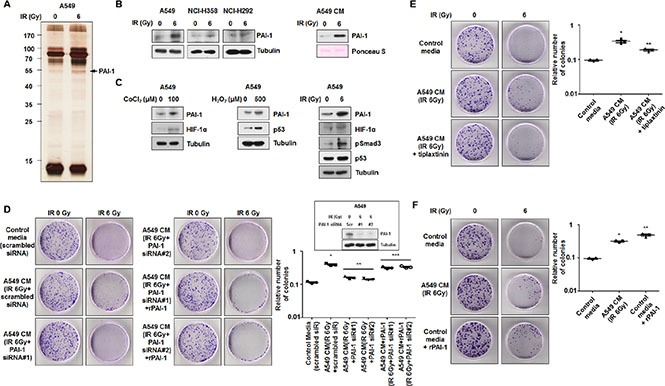
(A) The secretomes in CM of A549 cells exposed to 6 Gy were analyzed by silver staining and mass spectrometry. The band indicated by an arrowhead corresponds to PAI-1, which was increased by irradiation. (B) Radiation-induced expression levels of PAI-1 in A549, NCI-H358, and NCI-H292 cells and CM of A549 cells were analyzed by Western blotting. (C) Expression levels of PAI-1 and several transcription factors in A549 cells under hypoxia, ROS, or IR were analyzed by Western blotting. (D) The effects of PAI-1 knockdown on survival of NCI-H460 cells in response to radiation were measured by a colony forming assay using PAI-1-specific siRNA. The inset shows that siRNA oligonucleotides specific for PAI-1 significantly reduced PAI-1 expression in A549 cells measured by Western blotting. *p < 0.05 compared with irradiated cells treated with control media; **p < 0.05 compared with irradiated cells treated with CM of A549 cells without treatment of PAI-1 siRNA. (E) Effects of PAI-1 activation on survival of NCI-H460 cells in response to radiation were measured by a colony forming assay using tiplaxtinin (20 μM), a PAI-1 specific inhibitor. *p < 0.05 compared with irradiated cells treated with control media; **p < 0.05 compared with irradiated cells treated with CM of A549 cells not containing tiplaxtinin. (F) The effects of PAI-1 levels on survival of NCI-H460 cells in response to radiation were confirmed by a colony forming assay using rPAI-1 (50 ng/ml). *p < 0.05 compared with irradiated cells treated with control media; **p < 0.05 compared with irradiated cells treated with CM of A549 cells.