
Figure 4. Secreted extracellular PAI-1 stimulates downstream signaling in NCI-H460 cells by extracellular interaction, not by clathrin-mediated endocytosis.
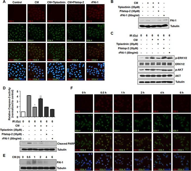
(A) Internalized PAI-1 in NCI-H460 cells after treatment with CM was analyzed by immunofluorescence. PAI-1, EEA-1, and cell nuclei are shown as red, green, and blue (DAPI) signals, respectively. PAI-1 endocytosed by clathrin-coated early endosome is indicated by co-stained red/green signals as yellow. (B) Intracellular levels of PAI-1 in NCI-H460 cells after treatment with CM were confirmed by Western blotting. (C) Effects of inhibition of PAI-1 endocytosis on AKT and ERK1/2 phosphorylation in NCI-H460 cells was analyzed by using tiplaxtinin (20 μM) or pitstop-2 (30 μM). (D) Effects of inhibition of PAI-1 endocytosis on activity of caspase-3 and PARP cleavage in NCI-H460 cells were analyzed using tiplaxtinin or pitstop-2. *p < 0.05 compared with irradiated cells treated with control media; **p < 0.05 compared with irradiated cells treated with CM. (E and F) Degradation of PAI-1 levels after endocytosis into NCI-H460 cells was analyzed by Western blotting and immunofluorescence, respectively. After treatment with CM from irradiated A549 cells, intracellular PAI-1 levels in NCI-H460 cells were measured from 0.5 h to 6 h.