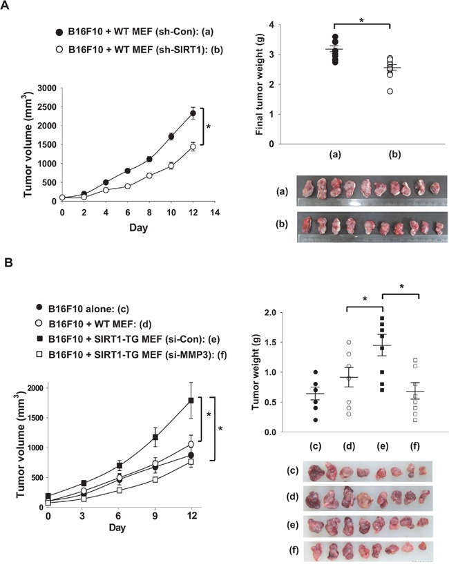
Figure 8. Fibroblast-derived MMP3 promotes tumor growth.

A. MEF cells obtained from WT mice were infected with sh-LTviral-Control or -SIRT1 (0.9 × 10⁸ TU/mL) for 72 hour. The MEF cells (1×10⁵) and B16F10 cells (1×10⁶) were mixed with Matrigel and injected into the flanks of nude mice. Tumor volumes are expressed at the means ± s.e.m. (n, 10; *, p < 0.05) in the left panel. Tumors (a, b) were excised and weighed on the final day. Each tumor weight is plotted in the right panel and pictures of tumors are shown below the plot. B. MEFs obtained from WT and SIRT1-TG mice were transfected with the indicated siRNAs (80 nM). MEFs (1×10⁵) and B16F10 cells (1×10⁶) were mixed with Matrigel and the cell mixtures were injected into the flanks of nude mice. Tumor volumes were measured from day 7 after implantation. Results are expressed as the means ± s.e.m. (n, 6-8; *, p < 0.05) in the left panel. Each tumor weight is plotted in the right panel and pictures of tumors are shown below the plot.
