
Figure 6. The phosphorylation of TOPK by Src enhances the stability of TOPK.
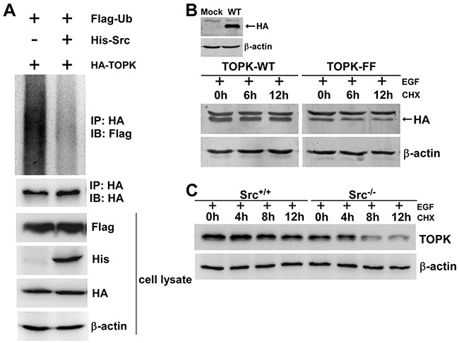
A. Phosphorylation of TOPK by Src prevented TOPK ubiquitination. HEK293T cells were transfected with pCMV-Flag-ubiquitin (Flag-Ub), pcDNA3-HA-TOPK (HA-TOPK), and pcDNA4-His-Src (His-Src) as indicated. The cells were harvested 48 h after transfection. Then the samples were immunoprecipitated (IP) with anti-HA and detected with anti-Flag by Western blot. The transfection efficiency and equal protein loading were verified by Western blot using the whole cell lysate. B. HEK293T cells were transfected with pcDNA3-HA-TOPK wild type (TOPK-WT) and pcDNA3-HA-TOPK double mutant (TOPK-FF), and then were treated with EGF (80 ng/ml; 30 min) followed by addition of CHX (100 μg/ml) to prevent new protein synthesis. The time-dependent stability of TOPK was detected with anti-HA by Western blot. C. Src+/+ and Src−/− MEFs were treated with EGF (20 ng/ml; 15 min) followed by addition of CHX (100 μg/ml) to prevent new protein synthesis. Lysates were collected at the indicated time points. The protein level of TOPK was detected by Western blot.