
Figure 7. Src phosphorylates TOPK to promote the tumorigenesis of colon cancer in vivo.
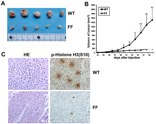
A. Tumors dissected from SW480/ WT or SW480/ FF group were shown. B. Tumor growth curve from mice injected with SW480/ WT or SW480/ FF cells. Data are expressed as means±SE of 5 mice in each group. The asterisk indicated a significant increase in tumor size in SW480/ WT injected mice compared with SW480/ FF injected mice. *P < 0.05, **P < 0.01. C. Left, H&E stained tumor sections from mice injected with either SW480/ WT or SW480/ FF cells. Poorly differentiated adenocarcinoma cell clusters were found in the two groups. Right, IHC analysis of p-Histone H3 (S10) expression in tumor sections from mice injected with either SW480/ WT or SW480/ FF cells. Nuclear expression of p-Histone H3 (S10) was detected in the tumor sections from SW480/ WT cells, while very weak staining of p-Histone H3 (S10) was detected from SW480/ FF cells. Magnification, 400×.