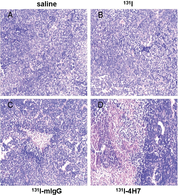
Figure 6. Samples dissected from renal cell carcinoma (RCC) cells treated with 131I-4H7, 131I-mIgG, 131I, or saline were sectioned and stained with hematoxylin-eosin for visualization of general morphology.
Representative images are shown (n = 8). Cells were treated with A. saline, B. 131I, C. 131I-mIgG, and D. 131I-4H7.

