Figure 1.

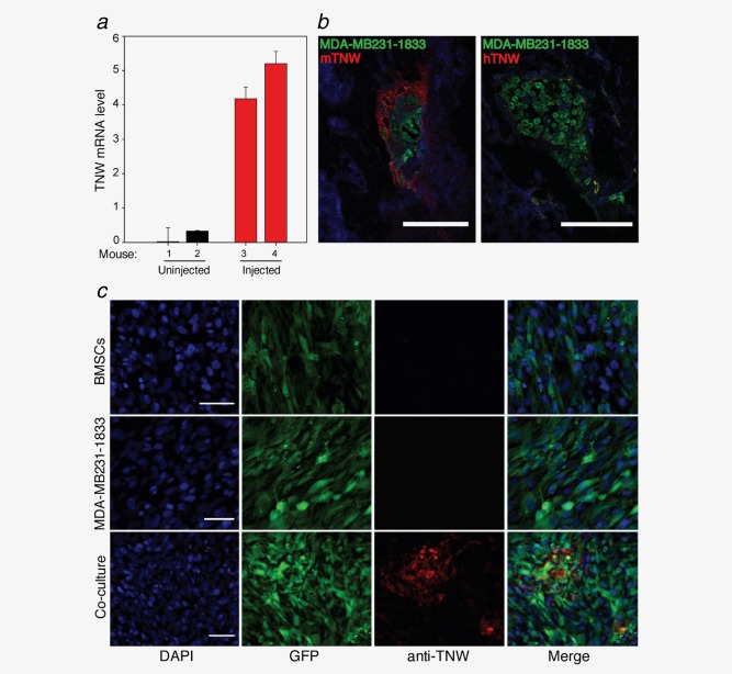
TNW is upregulated in bone metastasis of breast cancer. (a) TNW transcript levels in RNA isolated from osteoblasts sorted from two individual tumor‐free (1, 2, black) or two tumor‐bearing mice (3, 4, red). TNW levels relative to GAPDH were calculated using the relative standard curve method (ΔCt). Averages ± SD of two independent experiments in triplicate are shown. (b) Tissue sections of tibia show GFP‐MDA‐MB231‐1833 metastases (green). Staining of TNW was detectable with anti‐mouse TNW only (red, mTNW, left panel) but not with anti‐human TNW (hTNW, right panel). Scale bar, 100 µm. (c) Immunofluorescence staining for TNW of human bone marrow stromal cells (GFP‐BMSCs) and human breast cancer cells (GFP‐MDA‐MB231‐ 1833) cultured alone, or in co‐culture reveals TNW protein (red) expression after 7 days of co‐culture. Nuclei were labeled with DAPI (blue). Scale bars, 50 µm. [Color figure can be viewed in the online issue, which is available at wileyonlinelibrary.com.]
