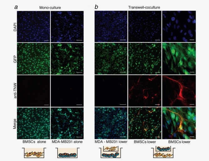
Figure 2.

Soluble factors secreted by MDA‐MB231‐1833 breast cancer cells stimulate TNW expression in BMSCs. (a) Monocultures of GFP‐BMSCs (left panels) and GFP‐MDA‐MB231‐1833 cells (right panels) maintained for 7 days in culture. Nuclei were stained with DAPI (blue). Staining with human anti‐TNW monoclonal antibody does not detect any TNW expression. (b) MDA‐MB231‐1833 cells or BMSCs were seeded in the bottom well of transwell chambers (MDA‐MB231‐1833 lower; BMSCs lower) and exposed to the other cell type in the upper chamber in an indirect co‐culture system. Under these conditions, TNW protein expression (red) was detected exclusively in BMSCs and not in MDA‐MB231‐1833 cells. Scale bars: 100 µm for all panels except for the magnification shown in the right panels representing 20 µm. [Color figure can be viewed in the online issue, which is available at wileyonlinelibrary.com.]
