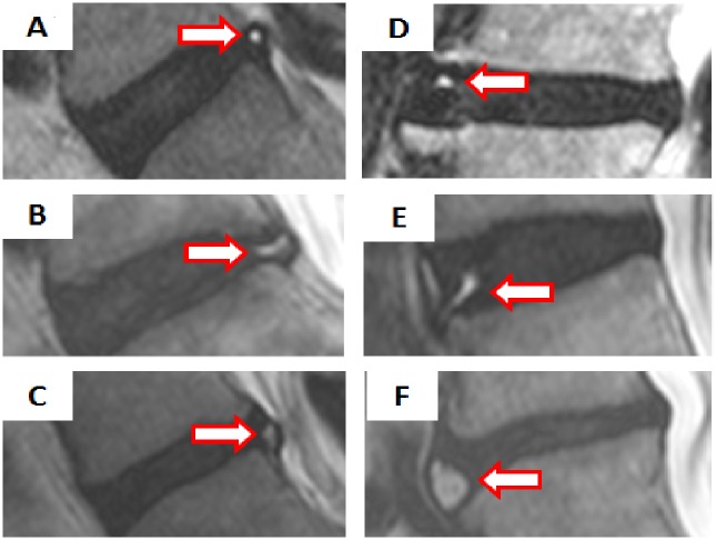
Fig 1. Classification of High Intensity Zones based on morphology and topography.
High Intensity Zones (HIZ) were defined as a high intensity signal (white) surrounded by low intensity (black) located in the annulus fibrosus on T2-weighted sagittal MRI. Six types of HIZs were created based on the shape (round type, fissure type, vertical type, rim type, and giant type), and location within the disc (posterior or anterior). The images represent (A) posterior round type, (B) posterior fissure type, (C) posterior vertical type, (D) anterior round type, (E) anterior rim type, and (F) anterior enlarged type.

