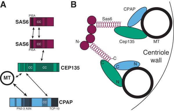
Figure 2. The interactions among core centriole proteins dictate the organization of the structure.
A) Known direct protein-protein interactions among core centriole proteins. Sas6 is included twice to illustrate its homotypic interactions. Shaded regions denote known or predicted protein motifs, PISA (present in Sas6), CC (coiled-coil), PN2-3 (a MT destabilizing motif), A5N (a MT binding motif) and TCP-10 (T complex protein 10). B) Cartoon of the proteins that compose 2 of the 9 arms of the cartwheel of a centriole and how they might interact to connect the symmetry of Sas6 to the MTs of the centriole. Approximate locations of the amino-terminal (N) and carboxy-terminal (C) ends of the proteins are indicated. See section 1.2 for details and references.

