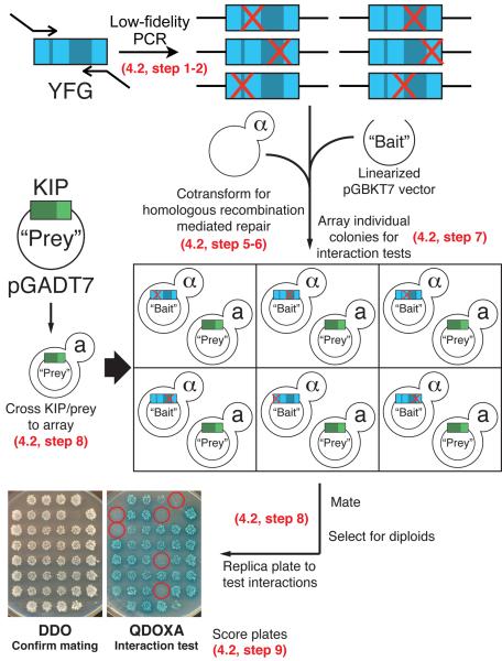
Figure 5. Workflow of an array based reverse Y2H screen to generate and identify mutations that disrupt protein-protein interactions.
Refer to sections indicated on the figure for details describing each step. Red X's represent random point mutations introduced by mutagenic PCR. Yeast colonies carrying mutations that disrupt the protein-protein interaction are circled in red.

