. 2016 Apr 29;8:ecurrents.md.77be6ec30e8caf19529a00417614a072. [Version 1] doi: 10.1371/currents.md.77be6ec30e8caf19529a00417614a072

Fig. 2. Myofibre necrosis in C57, untreated mdx and taurine treated mdx quadriceps muscle, from mice aged 22 days.
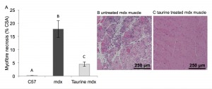
(A) Histological quantification of myofibre necrosis. Data are presented as mean ± SEM of percentage of cross section area (CSA) and n= 8 mice/group. Groups without a common letter are significantly (p<0.05) different. Representative images of myofibre necrosis and histology of H&E stained muscle sections are shown for (B) untreated mdx (C) taurine treated mdx mice.

(A) Histological quantification of myofibre necrosis. Data are presented as mean ± SEM of percentage of cross section area (CSA) and n= 8 mice/group. Groups without a common letter are significantly (p<0.05) different. Representative images of myofibre necrosis and histology of H&E stained muscle sections are shown for (B) untreated mdx (C) taurine treated mdx mice.