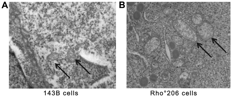
Figure 6.
A transmission electron microscopic comparison of 143B and Rho°206 cells. (A) ×20,000 magnification of 143B cells showing elongated mitochondria with parallel ordered cristae and normal electron density (indicated by black arrows). (B) ×20,000 magnification of Rho°206 cells. Mitochondria have an irregular cristae pattern and a 'whorled appearance' and are enlarged with almost complete or partial loss of the internal cristae structural pattern. Cytoplasmic vacuoles are enlarged and appear more numerous in (B) compared with (A).

