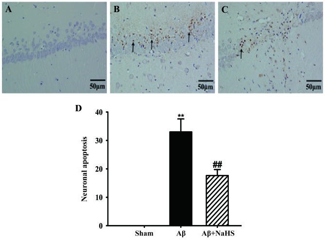
Figure 2.
Effect of sodium hydrosulfide (NaHS) on cell apoptosis in the hippocampal CA1 region of rats subjected to Aβ25–35-induced neurotoxicity by TUNEL staining in each group. (A) No apoptotic cells were observed in the hippocampus of rats from the sham-operated group. (B) Apoptotic cell death was increased in the Aβ25–35-exposed rat brains. (C) NaHS treatment significantly decreased the number of apoptotic cells. Arrows indicate apoptotic cells which were stained in brown. Scale bar, 50 µm. (D) Quantification of apoptotic cell in the hippocampal CA1 region. Data are presented as the means ± SEM (n=4 rats in each group, for triplicate experiments). **P<0.01 vs. sham-operated (sham) group ; ##P<0.01 vs. Aβ25–35 group.

