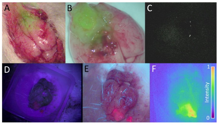
Fig. 4.
(A) The overlay (fusion) of two images acquired simultaneously during open craniotomy with the custom imaging module: the RGB white-light image and the fluorescence image overlay 2-h following a 3x microdose injection. (B) The ex vivo overlay of the whole brain acquired 2-h following a 6x microdose injection. (C) The corresponding ex vivo fluorescence image acquired with the Pentero IR800 channel. (D) A PpIX image acquired with the Zeiss Pentero BLUE400 channel, and (E) the same brain imaged by the custom imaging module during blue-light excitation. (F) The same brain as in D and E, imaged for ABY-029 fluorescence using the customized Pentero 2-h following a 3x microdose injection.

