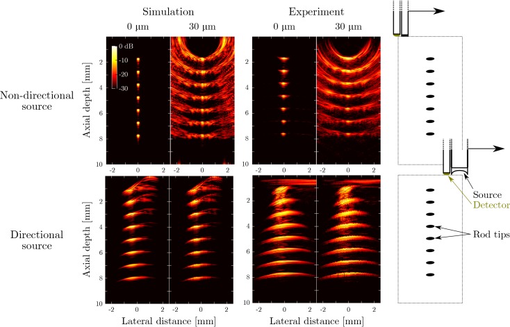
Fig. 3.
Top row: simulated (first two columns) and measured (middle two columns) all-optical ultrasound images obtained of a phantom using an unfocussed acoustic source. The phantom consisted of the tip of a single rod placed at different depths (right column); the resulting images are compounded into a single synthetic image. Simulations and experiments were performed both in the absence (“0 μm”) and presence (“30 μm”) of deliberate probe positioning errors. Positioning errors were applied to both the axial and the lateral axis and sampled from a uniform random distribution with a range of ±30 μm. Bottom row: the same panels are shown for the case where all-optical pulse-echo ultrasound data were acquired using the focussed acoustic source.

