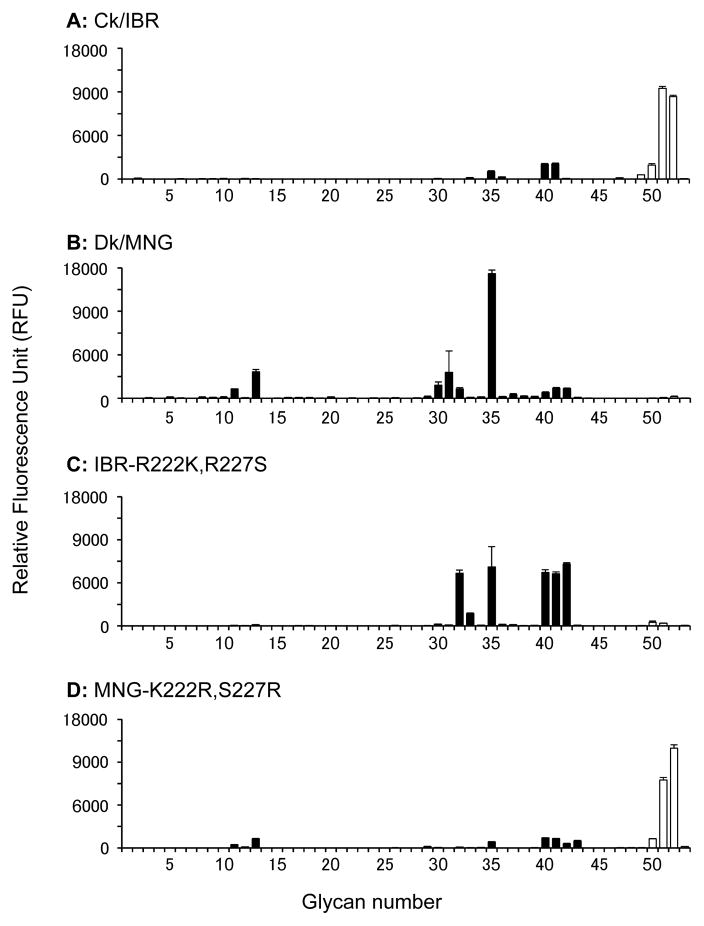
Fig. 2.
Glycan-binding specificity of the soluble trimeric recombinant hemagglutinins (rHAs). The glycan-binding specificity of rHA of Ck/IBR (A), Dk/MNG (B), IBR-R222K,R227S (C) and MNG-K222R,S227R (D) was analyzed by glycan microarray. Non-sialylated controls are shown as grey bars (#1–10), non-fucosylated α2,3 sialylated glycans are presented as black bars (#11–46) and fucosylated and α2,3 sialylated glycans are shown in white bars (#47–53). Each bar represents the mean signal minus background for each glycan sample and error bars are the SE value. Glycans imprinted on the array are listed in Table S1.

