
Figure 2. Btk Is Required for fMLF-Mediated Neutrophil Extravasation in the Murine Cremaster Muscle and Chemotaxis In Vitro.
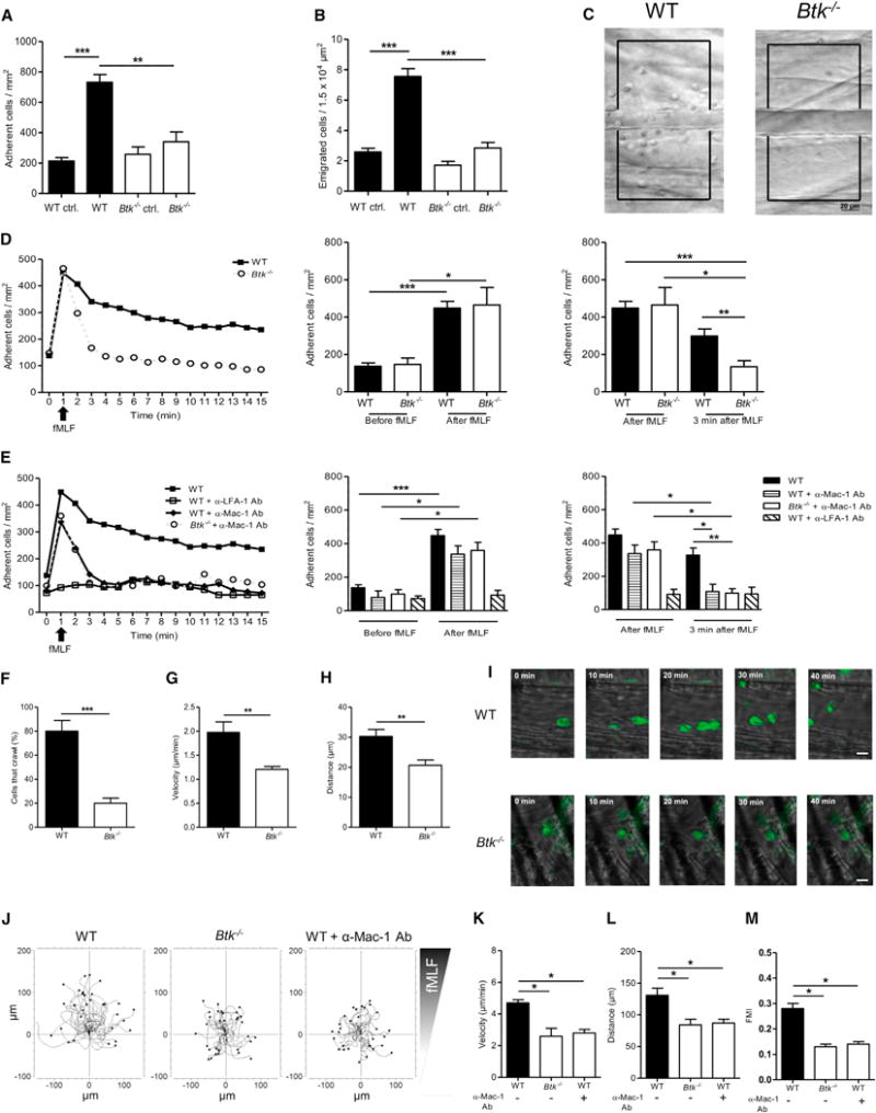
(A–H) Intravital microscopy of postcapillary venules in the murine cremaster was performed in WT and Btk−/− mice.
(A and B) Number of adherent cells per mm2 (A) and number of transmigrated cells per 1.5 × 104 μm2 (B) 4 hr after intrascrotal fMLF or vehicle injection.
(C) Representative images of inflamed WT (left) and Btk−/− (right) cremasteric venules visualized by reflected light oblique transillumination microscopy. Scale bar represents 20 μm.
(D and E) GPCR-induced arrest of neutrophils in postcapillary venules of WT (filled square) and Btk−/− (open circle) mice after fMLF injection (i.v.) (D) or after injection of blocking antibodies against LFA-1 (open square) or Mac-1 (filled diamond) prior to fMLF injection (E). Respective statistics are presented in related bar graphs.
(F–H) Intravascular crawling of Gr-1-labeled neutrophils during superfusion with fMLF. Percentage of adherent cells that crawled (F), crawling velocity (G), and crawled distance (H).
(I) Representative images of extravasated WT and Btk−/−. Scale bars represent 10 μm.
(J) Representative trajectory plots (30 cells from 3 independent experiments) of chemotaxing WT and Btk−/− neutrophils toward indicated fMLF gradient in vitro.
(K–M) Velocity (K), migration distance (L), and forward migration index (FMI) (M) of WT and Btk−/− neutrophils.
Depicted are mean + SEM; n ≥ 3 individual mice/group; *p < 0.05; **p < 0.01; ***p < 0.001 for all panels except (C), (I), and (J). See also Figures S2, S3, and S7 and Movies S3 and S4.