
Figure 7. Btk Is Involved in Integrin-Mediated Outside-In Signaling and FcRγ-Mediated Functions.
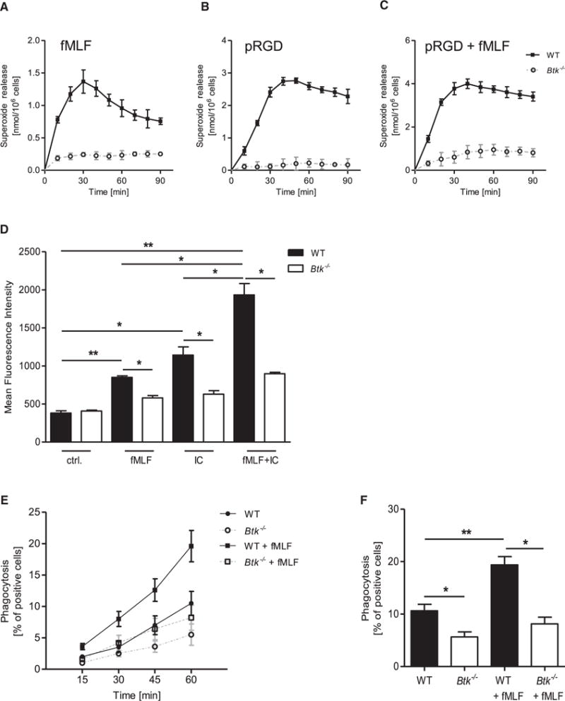
(A–C) Superoxide release of WT and Btk−/− neutrophils stimulated with 3 μM fMLF (A), plated on a polyvalent integrin ligand-coated surface (pRGD) without stimulus (B) or with 3 μM fMLF (C). Control values were subtracted from stimulated values.
(D) Quantitative binding of fibrinogen-coated fluorescent beads to unstimulated WT and Btk−/− neutrophils, stimulated with 1 μM fMLF, 10 μg/ml IgG immune complexes, or a combination of both as determined by flow cytometry.
(E) Phagocytosis of IgG-coated fluorescent beads by WT and Btk−/− neutrophils incubated at 37°C with or without 1 μM fMLF for indicated time points.
(F) Respective statistics of phagocytosis after 1 hr.
Depicted are mean + SEM of n ≥ 3 independent performed experiments; *p < 0.05; **p < 0.01; ***p < 0.001. See also Figure S7.