
Figure 5. Dally and Dlp depend on Prickle and Van Gogh.
Levels of Dlp were reduced in pk13 (A,B) and Vang6 (C,D) mutant clones (outlined with dashed white lines and lacking green fluorescence). (E–G) Levels of fluorescence in the wing discs of the protein trap Dally:YFP line were relatively uniform in controls (en-Gal4 UAS-RFP/+; dally:YFP/+), but were reduced specifically in the posterior compartment in the presence of pkRNAi (en-Gal4/+; dally:YFP/UAS-pkRNAi) and VangRNAi (en-Gal4/UAS-VangRNAi; dally:YFP/+). (H,I) Levels of Laminin detected by α-Laminin antibody staining (red) were reduced in pk13 mutant clones (outlined with dashed white line and lacking green fluorescence). Scale bar: 50 µm.
DOI: http://dx.doi.org/10.7554/eLife.18979.014
Figure 5—source data 1. Quantification of fluorescence intensity of α-Dlp staining, Dally:YFP and α-Laminin staining.
elife-18979-fig5-data1.xlsx (38.7KB, xlsx)
DOI: 10.7554/eLife.18979.015

Figure 5—figure supplement 1. Quantification of Dally and Dlp transcripts in Prickle and Van Gogh mutant wing discs.
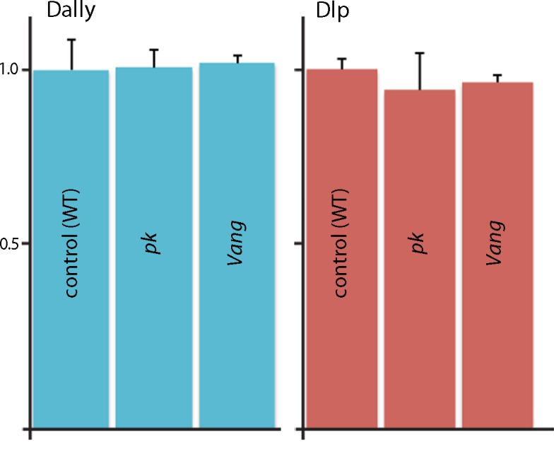
qRT-PCR was carried out to monitor expression of Dally and Dlp in normal, pk and Vang wing discs. Bar graph plots qRT-PCR for the Dally and Dlp transcripts relative to actin.
Figure 5—figure supplement 1—source data 1. Threshold cycles for dally, dlp and actin transcripts in wild-type, pk mutants and Vang mutants.
elife-18979-fig5-figsupp1-data1.xlsx (38.1KB, xlsx)
DOI: 10.7554/eLife.18979.017
Figure 5—figure supplement 2. Abnormal apical/basal polarity in Van Gogh mutant cells.

Image J-generated sagittal sections showing clones of Vang6 mutant cells (areas lacking GFP fluorescence and bounded by white dashed lines) had elevated levels of atypical Protein Kinase C (red, α–aPKC antibody staining), which is normally localized to the apical membrane domain and is downregulated by the PCP system. Discs large (α–Dlg antibody staining), which localizes basolaterally, was not apparently affected in the Vang6 mutant cells.