. 2016 Sep 20;5:e14810. doi: 10.7554/eLife.14810

Figure 6. Alfy localizes to axons and enriches to membrane fractions.

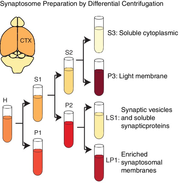
(A) Immunofluorescence images showing MAP2-positive neurons expressing Alfy. Merged color image demonstrates co-localization of mCherry-Alfy within a MAP2-positive neuron. Alfy is found within the soma and co-localizes with MAP2 positive projections. Transfections were replicated three times across three independent cultures. Colocalization to βIII Tubulin projections is shown in Figure 6—figure supplement 1. (B,C) Fractionation of adult cortical lysates reveals that Alfy enriches into membrane fractions. (B) Equal amounts of protein per fraction were analyzed by immunoblotting. Alfy was present in membrane fractions that also enriched with LC3-II and synaptophysin (P3, LP1; boxes. (C) The fold enrichment measured relative to the total homogenate fraction (H). Bars represent mean enrichment (n = 3) ± SEM. A schematic depiction of the fractionation can be found in Figure 6—figure supplement 2.

DOI: http://dx.doi.org/10.7554/eLife.14810.017

Figure 6.

Figure 6—figure supplement 1. Alfy is expressed in the soma and axons of neurons.

Figure 6—figure supplement 1.

(A,B) Alfy control mixed primary cortical cultures were transfected with plasmids containing full-length or truncated tagged-Alfy cDNA. (A) Representative confocal images are shown of Alfy-tagged constructs detected with antibodies directed against the fluorophore tag and co-stained MAP2 or BIII tubulin. Full-length and C-terminal fragments of Alfy are detected throughout MAP2- and BIII tubulin-positive projections (arrows). (B) Representative confocal images are shown of Alfy-tagged constructs detected with antibodies directed against the fluorophore tag (left) and co-stained with GFAP. Alfy are expressed throughout astroglia, although large vesicles and the nucleus are devoid of staining. Studies were replicated across three independent cultures. (C,D) To determine the sub-cellular distribution of Alfy, Alfy KO or Ctrl (A,B) mixed primary cortical cultures were transfected with plasmids containing full-length tagged-Alfy. To confirm transfection, COS-7 cells were transfected in parallel and immunoblotted. Vinculin is shown as a loading control. (C) Full-length Alfy-tagged proteins migrate at the expected size and are detected with anti-mCherry and anti-Alfy antibodies. Over-exposed images are necessary to show endogenous Alfy, since it is at a much lower abundance in non-neuronal cells (circle). (D) Full length and truncated constructs of Alfy. Studies were replicated across three cultures.
Figure 6—figure supplement 2. Alfy enriches in membrane fractions from brain.

Figure 6—figure supplement 2.

A schematic depiction of the modified synaptosome preparation used for Figure 6A, based on a previously published protocol (Hallett et al., 2008). Abbreviations: H, homogenate; P1, nuclei and large debris; S1, soluble cytoplasmic; P2, crude synaptosomal membranes; S2, soluble cytoplasmic; P3, light membrane; S3, soluble cytoplasmic; LP1, enriched synaptosomal membranes; LS1, synaptic vesicles and synaptic proteins.