. 2016 Aug 30;5:e16836. doi: 10.7554/eLife.16836

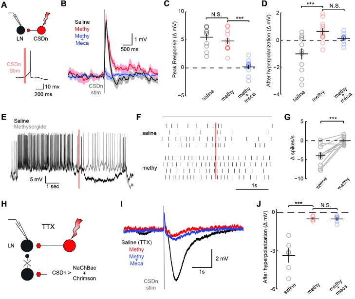
Figure 3. CSDn stimulation monosynaptically inhibits LNs and polysynaptically excites them.

(A) Schematic representation showing optogenetic stimulation of the CSDn and whole-cell recording of GABAergic LNs. bottom, stimulation of CSDn results in an action potential in an LNs. (B) LNs were held at −60 mV and CSDn stimulation resulted in a fast depolarization followed by a delayed hyperpolarization. Methysergide (50 μM, red) bocked the delayed hyperpolarization but has no effect on the depolarization. Mecamylamine (100 μM, blue) blocked the depolarization. (C,D). Summary statistics for B. Methysergide has no effect on the peak depolarizing response, n = 11, ANOVA, F = 58.93, p=4.13 × 10−9, saline versus methysergide p=0.42. The addition of mecamylamine eliminated the depolarization from CSDn stimulation, methysergide versus methysergide plus mecamylamine p=9.4 × 10−8. Methysergide did block the delayed hyperpolarization, ANOVA, F = 11.01, p=0.0006. Saline vs methysergide p=5.02 × 10−4. Mecamylamine had no effect the hyperpolarization p=0.3403. (E) An LN was depolarized to −30 mV to magnify the CSDn evoked inhibition. This inhibition is blocked by methysergide. (F) A raster plot showing the inhibition of LN spikes. Black horizontal line above raster denotes period of depolarization to −30 mV. CSDn stimulation occurred during the 40 ms red bar. (G) Summary of such experiments at −30 mV. n = 10, paired t-test p=8.47 × 10−4. (H) Schematic representation of NaChBac experiments. NaChBac and Chrimson are co-expressed in the CSDn. TTX is used to block all action potentials in the brain except in the CSDn. (I) NaChBac potentials in the CSDn cause a fast depolarization and delayed hyperpolarization in the LNs. The hyperpolarization is blocked by methysergide (50 μM, red). Mecamylamine (200 μM, blue) did not block the fast depolarization. (J) Summary statistics for experiments in I. ANOVA, n = 6, F = 44.25, p=1.08 × 10−5, saline vs methysergide p=2.70 × 10−5, methysergide vs methysergide plus mecamylamine p=0.999.

DOI: http://dx.doi.org/10.7554/eLife.16836.005

Figure 3.

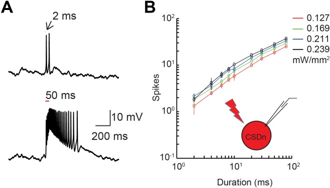
Figure 3—figure supplement 1. Demonstration and calibration of Chrimson activation of the CSDn.

Figure 3—figure supplement 1.

(A) top. A 2 ms flash of red light elicits approximately 2 action potentials from the CSDn, while a 50 ms stimulus a barrage of spikes (bottom). (B) Quantification of the relationship between light input and firing of the CSDn with Chrimson expression.
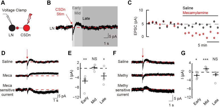
Figure 3—figure supplement 2. Stimulation of the CSDn depolarizes LNs via acetylcholinergic transmission and subsequently inhibits LNs via serotonin.

Figure 3—figure supplement 2.

(A) Schematic representation showing optogenetic stimulation of the CSDn and whole-cell voltage clamp recording of GABAergic LNs. (B) CSDn stimulation (red arrow) evokes dynamic responses in LNs. Red dashed line shows mean holding current prior to stimulation. Responses were divided into early, mid, and late responses. Data is the mean current from 19 LNs. (C) Time series of the peak amplitude of the early inward current in saline (black) and mecamylamine (100 μM, red). Data shown is mean of one experiment. (D) Mean current traces from LNs in response to CSDn stimulation across six flies. Subtraction of currents in mecamylamine (Meca, middle) from saline-recorded currents (top) reveals the total current that is sensitive to mecamylamine (bottom). (E) Summary plot for the early, mid, and late phase mecamylamine sensitive currents. Early = p=0.004, Mid = p=0.507, Late = p=0.034, n = 6, one-sample t-test. (F) Mean current traces from seven flies in saline (top) and methysergide (methy, middle). The methysergide-sensitive current is shown on bottom. (G) Summary plot for the early, mid, and late phase methysergide-sensitive currents. Early = p=0.032, Mid = p=0.0007, Late = p=0.128, n = 7, one-sample t-test.
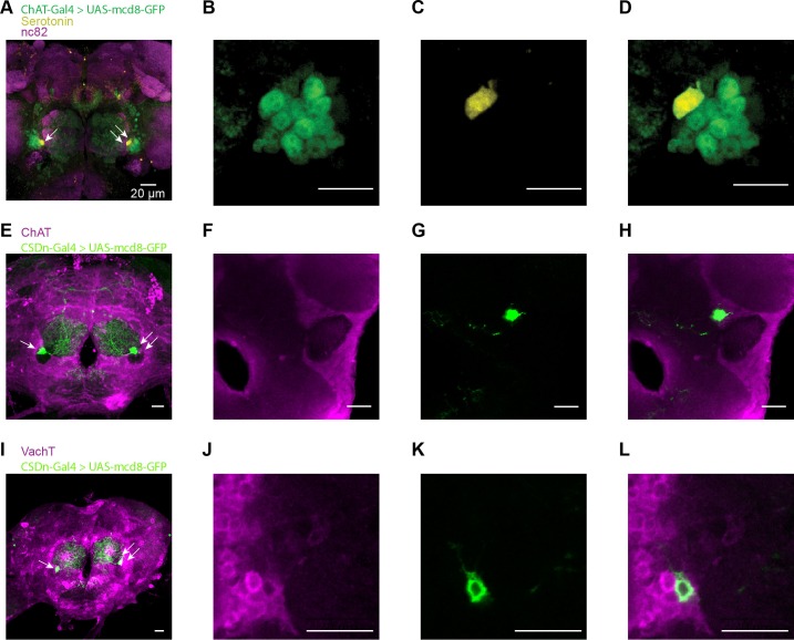
Figure 3—figure supplement 3. The CSDn may release acetylcholine as a co-transmitter.

Figure 3—figure supplement 3.

(A) Membrane-bound GFP is expressed via the promoter, ChAT, to reveal cholinergic neurons and its co-localization with the 5-HT antibody. The CSDn is identified as the sole 5-HT positive neuron adjacent to the antennal lobe. nc82 = magenta, mcd8-GFP = green, and 5-HT = yellow. Scale bar = 20 μm for all panels. (BD) A single slice of the brain is shown at the level of the CSDn with GFP and 5-HT labeling. (E) The CSDn is labeled with mcd8-GFP and the brain is stained with a ChAT antibody. (FH) A single slice through the brain at the level of the CSDn reveals that it is labeled with the ChAT antibody. (I) The CSDn is labeled with mcd8-GFP and the brain is stained with the VAchT antibody.(JL) A single slice through the brain at the level of the CSDn reveals that it is labeled with the VAchT antibody.
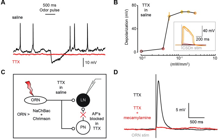
Figure 3—figure supplement 4. Co-expression of the TTX-insensitive sodium channel, NaChBac, and Chrimson can be used to effectively test mono-synaptic versus poly-synaptic connections.

Figure 3—figure supplement 4.

(A) A recording from a CSDn that co-expresses NaChBac and Chrimson. The CSDn displays normal action potentials and is inbibited by the presentation of odors (black trace). The addition of TTX (1 μM) to the saline bath blocks all typical firing in the CSDn and blocks the odor-evoked inhibtion (red trace). This is presumably due to the prevention of action potentials in ORNs and LNs. (B) Red light drives subthreshold activity in the CSDn via Chrimson expression even in TTX. Higher levels of Chrimson activation result in a broad NaChBac-mediated plateau potential. (C) Design of a proof-of-principle experiment to demonstrate that NaChBac plateau potentials can mediate synaptic transmission. NaChBac and Chrimson are co-expressed in ORNs using the Orco-Gal4 promoter and an LN in the AL is patched. TTX is used to block all activity in the brain, thus blocking all poly-synaptic contributions. The ORNS are stimulated with Chrimson and ORN APs are mediated by NaChBac. (D) Chrimson stimulation of NaChBac-expressing ORNs results in a large post-synaptic depolarization in an LN even in the presence of TTX. TTX blocked all action potentials in the LN both at rest and during ORN stimulation. This connection is blocked by the acetylcholine antagonist mecamylamine (200 μM).