Figure 2.
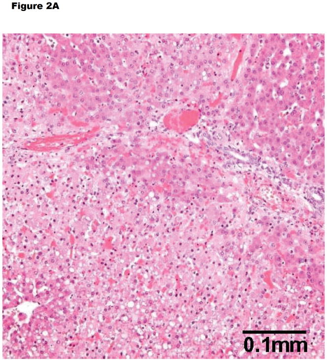
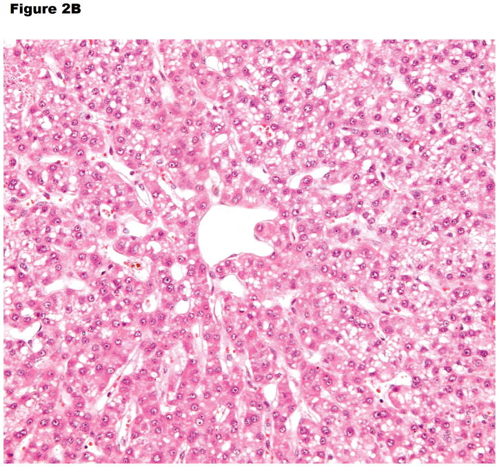
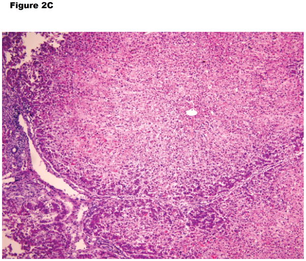
Histopathology of (A) hyperacute rejection (<24 hours) in a wild-type pig liver transplanted orthotopically into a baboon, (B) a GTKO/hCD46 orthotopic pig liver graft in a baboon that survived for 6 days, and (C) a pig left liver lobe graft in a Tibetan monkey that survived for 14 days
(A) WT pig-to-baboon liver xenotransplantation at 1 h (x200). Severe hepatocellular vacuolar change, focal hepatocyte necrosis, and few thrombi.
(B) Vacuolar hepatocellular cytoplasmic change with minimal hepatocellular necrosis on postoperative day 6 (x200).
(C) The graft shows some lymphocyte infiltration in the portal area, but no major features of antibody-mediated or cellular rejection (x100).


