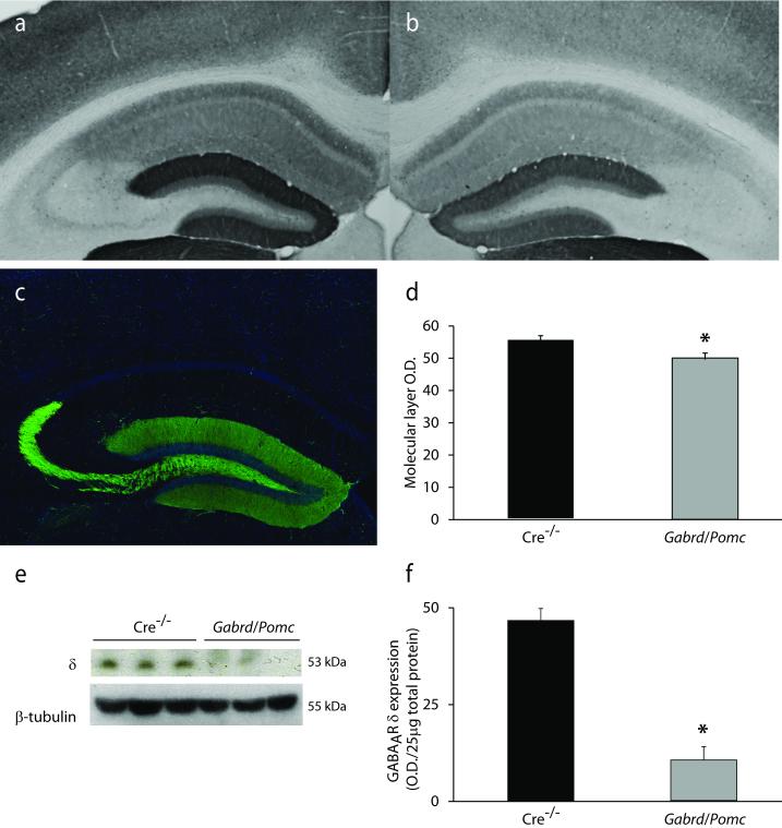
Figure 5. Reduced expression of the GABAAR δ subunit in the dentate gyrus of Gabrd/Pomc mice.
Representative images of immunoreactivity for the GABAAR δ subunit in Cre−/− littermates (a) and Gabrd/Pomc mice (b). c, Representative image of GFP reporter expression in Pomc-GFP mice, demonstrating the specificity of Cre expression in dentate gyrus granule cells. d, The average optical density of GABAAR δ subunit immunoreactivity is decreased in Gabrd/Pomc mice compared to Cre−/− littermates. n = 3 mice per experimental group. e, Representative Western blot of GABAAR δ subunit expression in Gabrd/Pomc mice and Cre−/− littermates. f, The average optical density of GABAAR δ subunit expression, quanitified using Western blot analysis, is decreased in Gabrd/Pomc mice Gabrd/Pomc mice compared to Cre−/− littermates. n = 6 mice per experimental group; * denotes significance of p<0.05 using a Student’s t-test.

