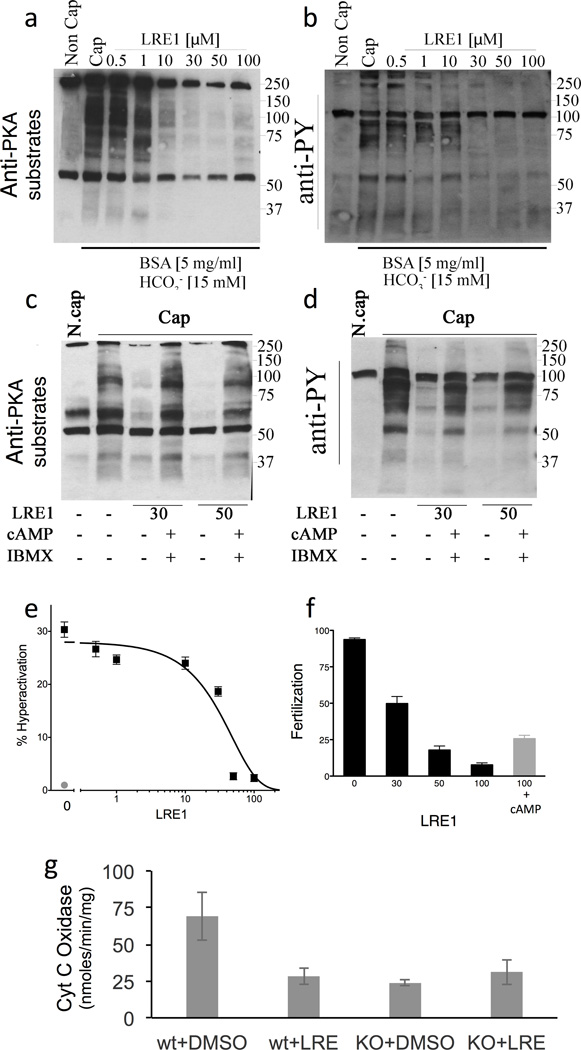
Fig. 5. LRE1 inhibits sAC dependent processes in sperm and mitochondria.
(a–d) Sperm experiments. (a) Western blot using anti-PKA substrates antibodies of mouse cauda sperm activated by incubation in capacitation (Cap) media in the presence of the shown amounts of LRE1 in presence (+) or absence (−) of dibutyryl cAMP (1 mM) and IBMX (100 µM). N.Cap = non capacitated negative control. (b) Western blot using anti-phospho tyrosine antibodies of the same blot as in (a). For a,b, shown are representative Western blots of experiments repeated three times using independent sperm preparations from different mice. The complete gels used for these images are shown in Supplementary Figures 4 and 5, respectively. (c) The percentage of hyperactivated motility in sperm capacitated in the presence of the indicated concentration of LRE1 (black). Non capacitated sperm (red). Values are averages (± S.E.M.) of three independent sperm preparations from three different mice collected and analyzed on separate days. (d) Percentage of fertilized eggs from sperm capacitated in the presence of the indicated concentration of LRE1 (black bars) or sperm capacitated in the presence of 100 µM LRE1 + 1 mM dibutyryl cAMP (red bar). Values are averages (± S.E.M.) of four independent sperm preparations from four different mice collected and analyzed on separate days. (e) Cytochrome c oxidase (COX) activities in cells from WT and sAC KO MEFs treated with DMSO or 50 µM LRE1 for 30 min. Values are averages (± S.D.); N=5. COX activity was statistically different in WT cells ± LRE1 (P=0.037) by t-test.

