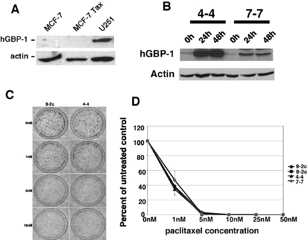
Fig. 1.
A. hGBP-1 was not expressed in paclitaxel-sensitive and -resistant MCF-7 cells. U251 glioblastoma cells were used as a positive antibody control. B. Control vector (8-2c and 9-2c) and tetracycline (tet)-regulated MCF-7 cells (4-4 and 7-7) were incubated in the absence of tet for the times indicated and examined for hGBP-1. C. Tet-regulated control and hGBP-1-expressing cells were treated with the concentrations of paclitaxel listed for 48 h. Drug was removed and the cells incubated in media without tet for 7–10 days. Colonies were visualized by staining with crystal violet. Representative photomicrographs are shown. D. The numbers of colonies with 50 or more cells were counted. Results of three experiments performed in triplicate are shown. Results are normalized to the number of colonies generated by untreated control cells. The average number of colonies ± SD are shown (n = 3).

