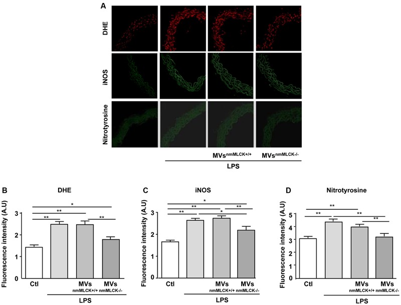
FIGURE 4.

Microvesicles from non-muscular myosin light chain kinase-deficient mice (MVsnmMLCK-/-) reduce the LPS-evoked oxidative and nitrative stresses in mouse aorta. (A) Confocal image staining of DHE (red), iNOS (green), and nitrotyrosine expression (green) in mouse wild type aorta exposed 24 h ex vivo to saline salt solution, LPS alone (10 μg/ml), LPS + MVsnmMLCK+/+ or LPS + MVsnmMLCK-/-. Aorta was imaged using confocal microscope. (B–D) Histograms show fluorescence intensity of aorta DHE staining (B), iNOS (C), and nitrotyrosine (D) assessed by Image J. Data represent arbitrary units (A.U.) of the mean ± SEM (n = 3–5). ∗P < 0.05, ∗∗P < 0.01.
