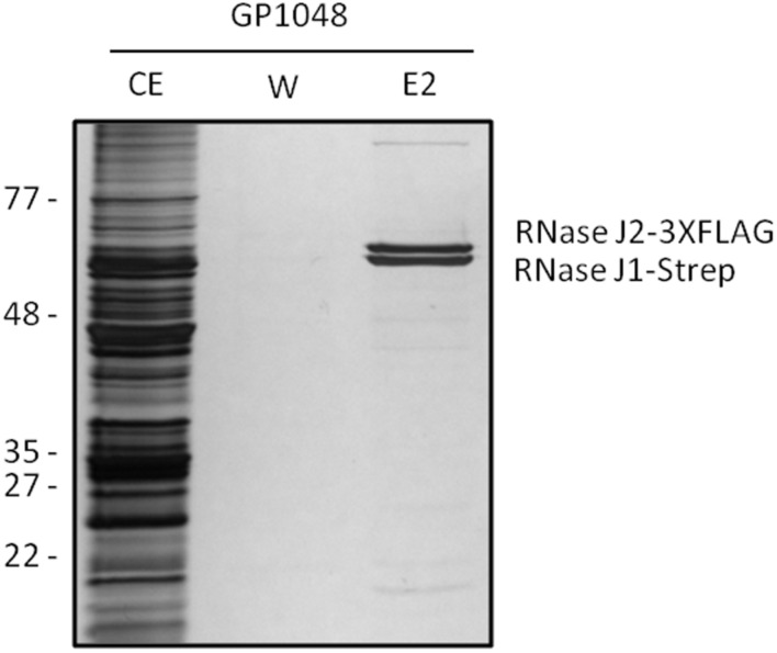
FIGURE 2.
RNases J1 and J2 form a complex in vivo. Bacillus subtilis GP1048 was cultivated in LB medium at 37°C to OD600 of 1.0. Cells were disrupted and Strep-RNase J1 was purified by a StrepTactin column. CE, cell extract; W, wash fraction; E2, elution fraction 2. The identity of RNases J1 and J2 was proven by Western blot analysis (data not shown). The faint eluted bands at the top and the bottom of the gel correspond to PycA and AccB, respectively, the two biotin-containing proteins of B. subtilis (Meyer et al., 2011).

