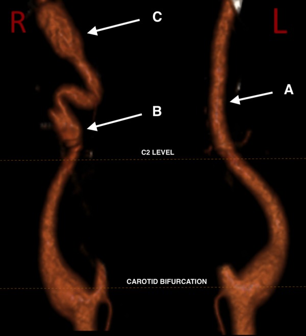
Figure 3.

Volume-rendered CT angiogram demonstrating complete remodelling on the left (A) but persisting irregularity on the right with a shallow dissecting aneurysm at the C1/2 level (B) and a larger, saccular dissecting aneurysm at the skull base (C).
