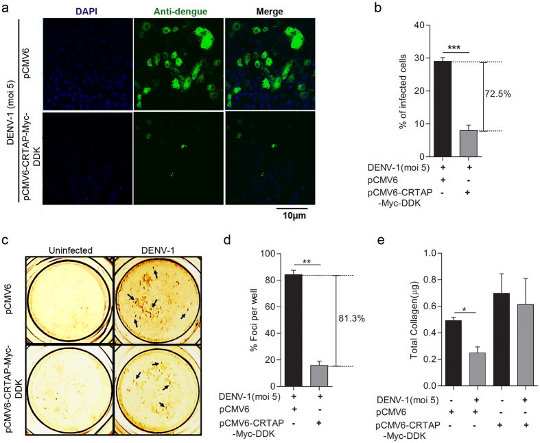
Figure 6. Over-expression of CRTAP gene inversely regulates DENV-1 infection.
(a) Laser scanning confocal microscopy to visualize the number of DENV-1 infected Vero cells transfected with either pCMV6 (vector control) or pCMV6-CRTAP-Myc-DDK. Vero cells were transfected with equal concentrations of both the plasmids for 36 hours followed by infection of DENV-1 (moi 5) for another 18 hrs. Following incubation, the cells were fixed, permeabilized, and stained with anti-Dengue monoclonal Ab (green). (b) Quantification of the number of DENV-1 infected cells in vector transfected and pCMV6-CRTAP-Myc-DDK transfected Vero cells. Infected cells were counted as mentioned in the previous figure. (c) Foci forming unit reduction assay (FFURA) on vector transfected and pCMV6-CRTAP-Myc-DDK transfected Vero cells was performed to determine the antiviral activity of CRTAP on DENV-1 as already described in material and methods. Arrows indicate representative DENV-1 foci. (d) Data from duplicate assays of three independent experiments were plotted. The percentage of foci reduction is represented in the graph. (e) Collagen estimation was done in vector transfected or pCMV6-CRTAP-Myc-DDK transfected cell lysates as described in material and methods. DENV-1 infection was given as indicated. Statistical analysis was performed using Student’s t-test using Graph Pad Prism version 5 (Graph Pad Software Inc., San Diego, CA.). Error bars represent the standard error of the mean (SEM). ***P < 0.001, **P < 0.01, *P < 0.05 were considered statistically significant.

