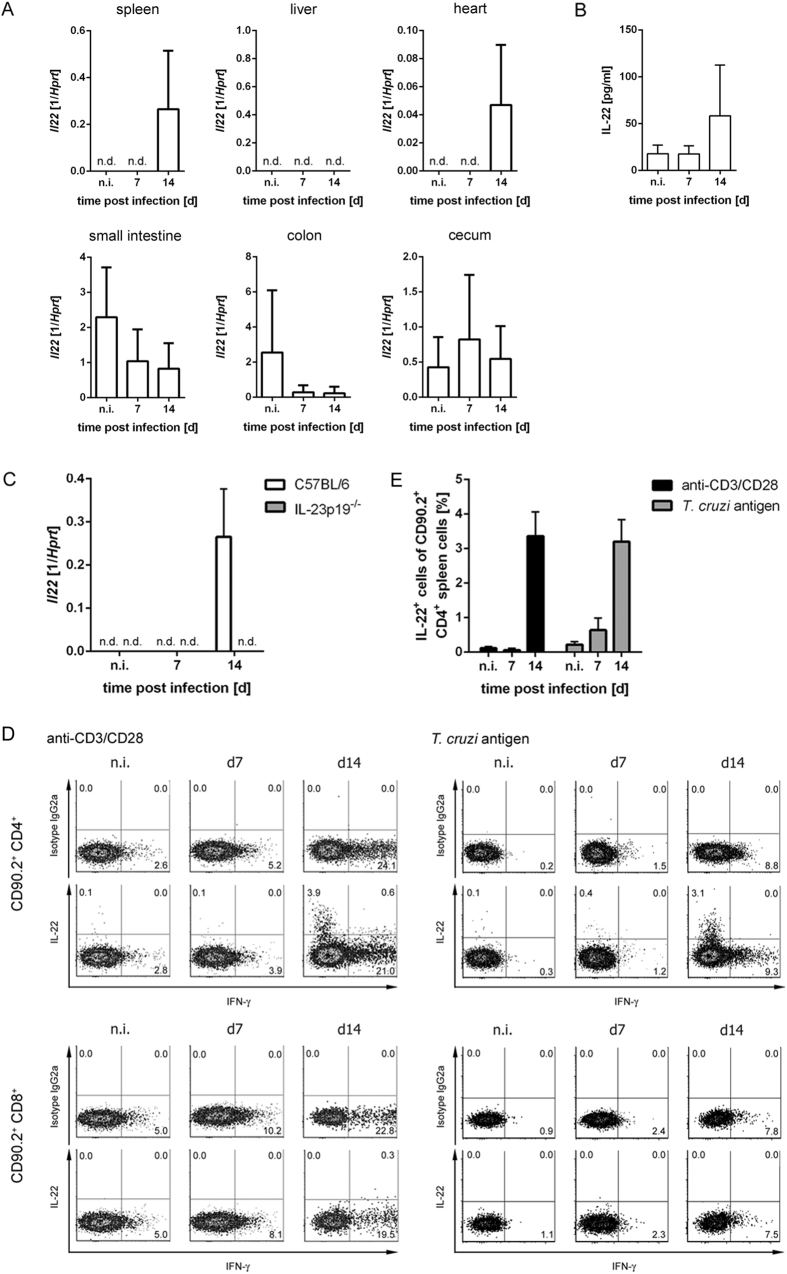
Figure 1. IL-22 is produced in response to T. cruzi infection.
C57BL/6 mice were infected i.p. with 500 T. cruzi trypomastigotes. (A) Il22 mRNA expression in the spleen, liver, heart, small intestine, colon and cecum was quantified by real-time PCR. (B) The concentration of IL-22 in the sera was determined by cytometric bead array. (C) C57BL/6 mice and IL-23p19−/− mice were infected i.p. with 500 T. cruzi trypomastigotes and the Il22 mRNA expression in spleens was quantified by real-time PCR at the indicated time points post infection. Results are expressed as means ± standard deviations of 4–5 mice per group. (D) The intracellular IL-22 and IFN-γ production of C57BL/6 spleen cells was analyzed by flow cytometry after anti-CD3/CD28 stimulation or specific stimulation with T. cruzi antigen. Cells were gated on CD90.2+ CD4+ or CD90.2+ CD8+ T cells, respectively. Shown are representative dot plots of from 5 mice per group. (E) Percentages of IL-22-producing cells out of CD90.2+ CD4+ T cells after anti-CD3/CD28 stimulation or after antigen-specific stimulation (as shown in (D)). Results are expressed as means ± standard deviations of 5 mice per group. n.d. not detectable; n.i. not infected.

