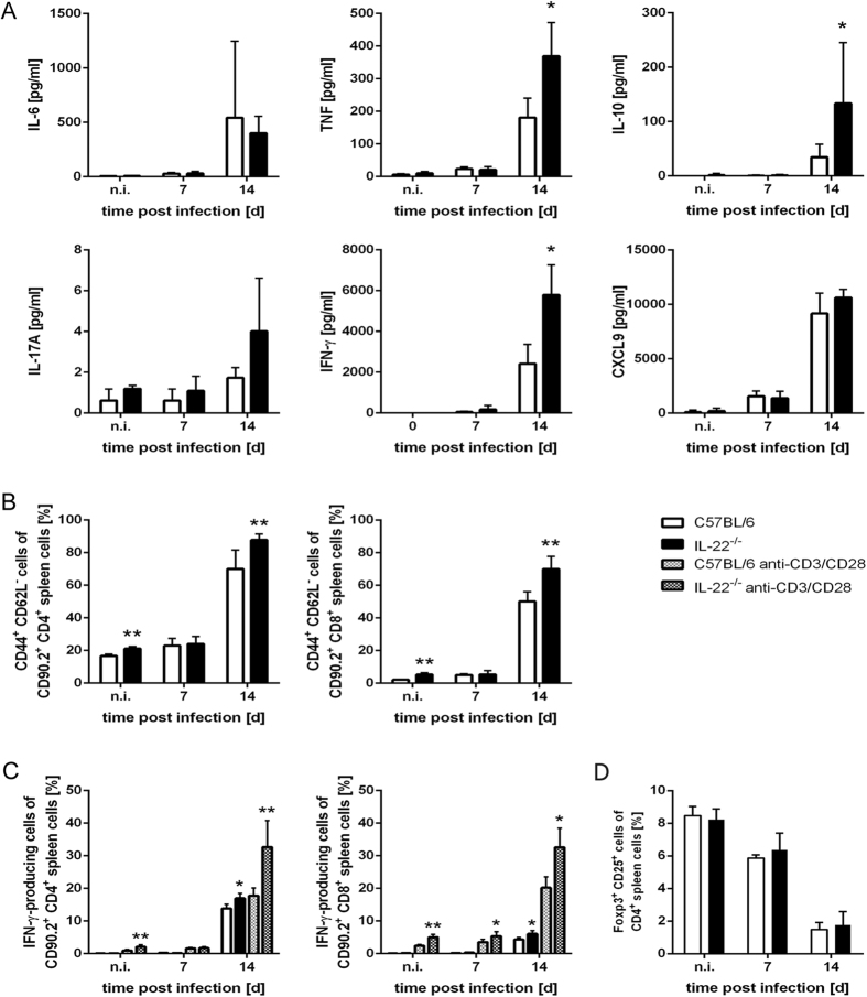
Figure 2. Increased cytokine and chemokine production as well as enhanced numbers of activated T cells in IL-22−/− mice during T. cruzi infection.
C57BL/6 mice and IL-22−/− mice were infected i.p. with 500 T. cruzi trypomastigotes. (A) Single cell suspensions of spleen cells were cultured for 24 h and the secretion of different cytokines and chemokines into the supernatant was determined by cytometric bead array. (B) Spleen cells were analyzed by flow cytometry at the indicated time points after infection. Shown is the percentage of activated (CD44+ CD62L−) cells of CD90.2+ CD4+ or CD90.2+ CD8+ spleen cells, respectively. (C) Spleen cells were stimulated with anti-CD3/CD28 or left unstimulated and were analyzed for intracellular IFN-γ production. Shown is the percentage of IFN-γ-producing cells of CD90.2+ CD4+ or CD90.2+ CD8+ spleen cells, respectively. (D) The percentage of Foxp3+ CD25+ Tregs out of CD4+ T cells was analyzed by flow cytometry. Results are expressed as means ± standard deviations of 5 mice per group. * and ** indicate statistical significance (p ≤ 0.05 and p ≤ 0.01, respectively) compared to C57BL/6 wild-type mice.

