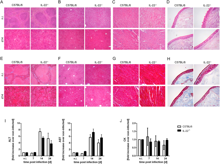
Figure 6. Unaltered pathology in T. cruzi-infected IL-22−/− mice.
C57BL/6 mice and IL-22−/− mice were infected i.p. with 500 T. cruzi trypomastigotes. Tissue sections of spleen (A,E), liver (B,F), heart (C,G) and colon (D,H) were stained with H&E (A–D) or with azan trichrome (E–H). Representative sections from five mice per group (2 uninfected mice per group) are shown. Magnification: spleen, liver, colon 100x; heart 200x. Liver derived enzymes ALT and AST (I) and CK (J) were quantified in sera at the indicated time points after infection. Results are expressed as means ± standard deviations of 5 mice per group (2–5 uninfected mice per group). Similar results were obtained in two independent experiments.

