Figure 4.
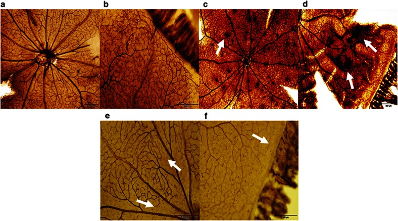
Retinal flatmounts of P21 RA controls, Sal/Sal IHR, and Keto/Caff-treated IHR retinas. Panels a and b represent RA controls, panels c and d represent Sal/Sal IHR groups, and panels e and f represent Caff/Keto-treated IHR groups. RA images show normal retinal vasculature in the optic disk (panel a) and periphery (panel b). The placebo-treated IHR groups show punctate hemorrhages at the optic disk (panel c, arrow), and neonvascularization, vascular tufts, and tortuous, dilated vessels consistent with severe OIR at the periphery (panel d, arrows). Caff/Keto reduced the severity of OIR with evidence of capillary dropout at the optic disk (panel e, arrow) and periphery (panel f, arrows). Images are 4× magnification, bar scale is 200 µm.

