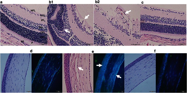
Figure 5.
H&E and COX-2 immunofluorescence of retinal layers from P21 RA controls, Sal/Sal IHR, and Keto/Caff-treated IHR groups. Panels a and d represent RA controls, panels b1, b2 and e represent Sal/Sal IHR groups, and panels c and f represent Caff/Keto-treated IHR groups. RA control retina and cornea appear normal. Sal/Sal IHR retinas show neovascularization retinal cells invading the vitreous fluid (panel b1, 40× and panel b2, 60×, arrows), retinal folding, and photoreceptor abnormalities (panel b1, arrows), as well as corneal degeneration with increased COX-2 immunostaining (e, arrows). Treatment with Caff/Keto reduced the effects (panels c and f). Bar scale is 20 µm. NFL (nerve fiber layer); GCL (ganglion cell layer); IPL (inner plexiform layer); INL (inner nuclear layer); OPL (outer plexiform layer), ONL (outer nuclear layer); P (photoreceptors); PE (pigmented epithelium); C (choroid).

