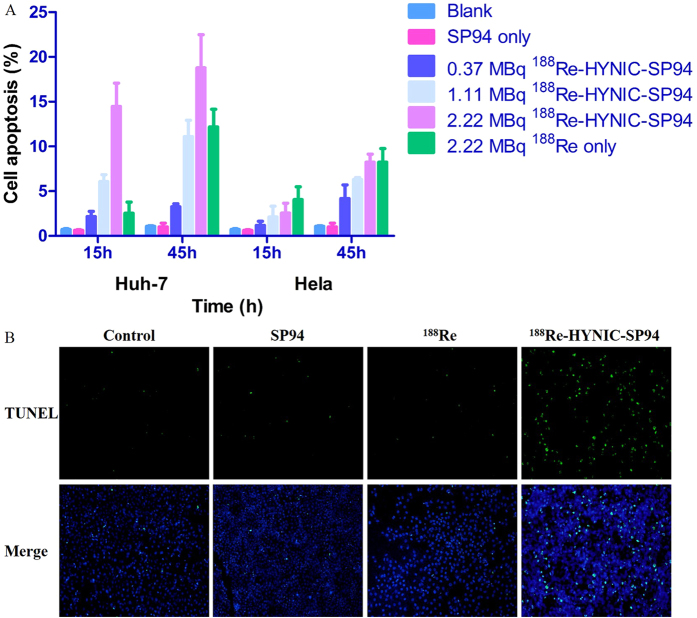
Figure 5. Comparison of different conditions induced apoptosis in Huh-7 and Hela cells by TUNEL assay.
(A) Quantitative analysis of apoptosis in Huh-7 and Hela cells. (B) Images of captured apoptotic cells by a fluorescence microscope; the green-fluorescent signal represented cells that underwent apoptosis and DAPI was used for a nuclear stain.

