
Figure 1.
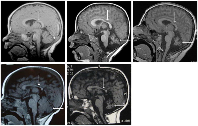
All images are T1-weighted sagittal MRIs. Images (a–c) are of Patient 1 at 11 months old (a), 3.6 years old (b), and 12.5 years old (c). Image (a) shows an age-appropriate size of the cerebellum. Images (b) and (d) illustrate cerebellar atrophy that progresses with age. All three images show thinning of the body of corpus callosum, particularly in the posterior aspect. Images (d–e) are of Patient 2 at 6 months old (a) and 6 years old (e). Again, image (d) shows an age-appropriate cerebellar size, whereas image (e) illustrates cerebellar atrophy with time. Thinning of the corpus callosum is also apparent in (d) and (e). The corpus callosum is designated by a vertical arrow and the cerebellum by a horizontal arrow.