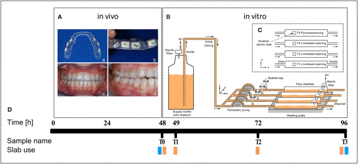
Figure 1.
Experimental outline. (A) Dental splint with six human enamel-dentin slabs (white squares, a, top view; b, side view) fixed in the upper jaw (c, front view; d, side view). (B) Sketch of the biofilm reactor setup (from left to right): Supply bottle filled with BHI medium (orange), peristaltic pump, bubble trap, DFR 110 biofilm reactor on heating plate (34°C), arrows indicate medium flow direction. (C) Top view sketch of the enamel-dentin slab distribution in biofilm reactor chambers. (D) Timeline and slab use (orange, live/dead staining; blue, pyrosequencing).

