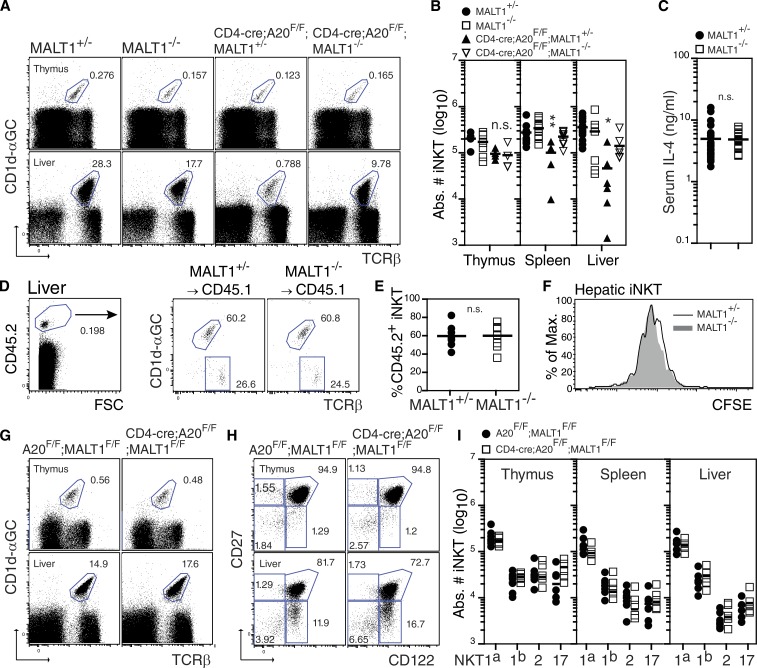
Figure 4.
MALT1 deficiency rescues iNKT cell development in CD4-cre;A20F/F mice. (A) Frequency and absolute iNKT cell counts (B) in thymus and liver of MALT1+/− (n = 13), MALT1−/− (n = 12), CD4-cre;A20F/F;MALT1+/− (n = 6), and CD4-cre;A20F/F;MALT1−/− (n = 7) mice, respectively. Data are pooled from four independent experiments. (C) Serum IL-4 concentration in MALT1+/− (n = 26) and MALT−/− (n = 20) mice after i.p. injection of α-GalCer. Results are pooled from two independent experiments. (D and E) Frequency of MALT1+/− (n = 4) and MALT1−/− (n = 4) iNKT cells recovered from CD45.1 congenic mice after transfer of CFSE-labeled CD8-depleted thymocytes. (F) Homeostatic proliferation of MALT1+/− and MALT1−/− iNKT cells in the livers of congenic mice 8 d after transfer. Data are from two independent experiments pooled. (G–I) Frequency and absolute iNKT cell counts in thymus, spleen, and liver of control A20F/F;MALT1F/F (n = 6) and CD4-cre;A20F/F;MALT1F/F (n = 6) mice, respectively. FACS dot blots in H are gated on βTCR+CD1d-αGC+ iNKT cells from each organ. Data are pooled from three independent experiments.*, P < 0.05; **, P < 0.01. n.s., not significant.

