Figure 5.
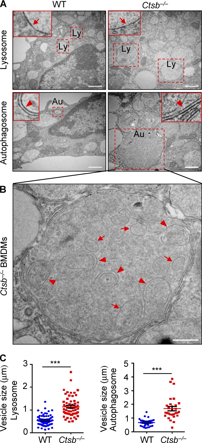
Cathepsin B deficiency results in impaired lysosomal recycling. (A) Transmission electron microscopy analysis of WT and Ctsb−/− BMDMs. Arrows in each inset indicate the single membrane lysosome– or double membrane autophagosome–associated vesicles. Dashed squares indicate a lysosome (Ly) or autophagosome (Au). (B) Undigested vesicles are accumulated in the autolysosomes of Ctsb−/− BMDMs. Arrows indicate double membrane–enclosed vesicles, and arrowheads indicate single membrane–enclosed vesicles. (C) Quantification of the size of lysosomes and autophagosomes from WT and Ctsb−/− BMDMs. The diameters of more than 60 lysosomes and 30 autophagosomes were analyzed. Data are representative of two independent experiments and are means ± SEM. Bars: (A) 1 µm; (B) 0.5 µm. ***, P < 0.001.

