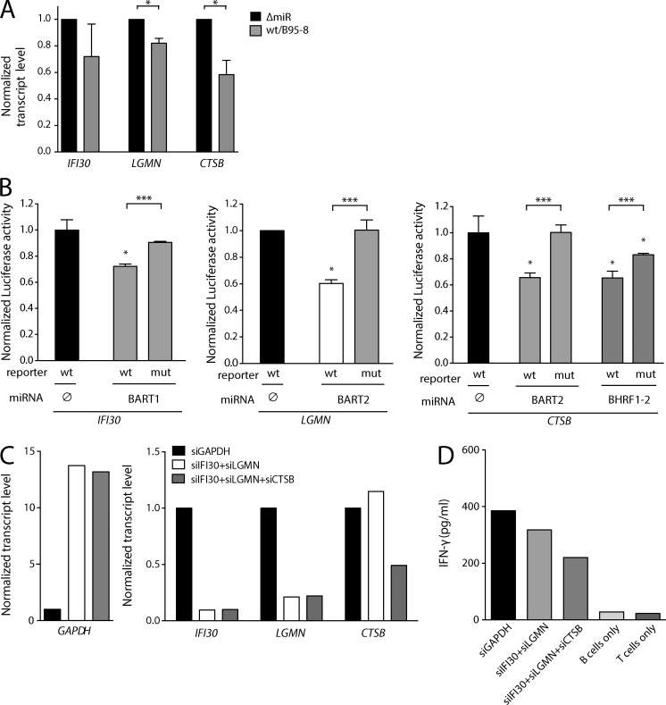
Figure 5.
Viral miRNAs inhibit two lysosomal endopeptidases and a thiol reductase needed for antigen presentation. (A) Transcript levels of IFI30, LGMN, and CTSB encoding GILT, AEP, and CTSB, respectively, were measured with quantitative RT-PCR (n = 3). P-values were calculated by a paired two-tailed Student’s t test. *, P < 0.05. (B) HEK293T cells were cotransfected with different miRNA expression vectors and luciferase reporter plasmids carrying 3′-UTRs as indicated (n = 3). The luciferase activities were normalized to lysates from cells cotransfected with the wild-type 3′-UTR reporter and an empty plasmid. wt, wild-type 3′-UTR; mut, mutated 3′-UTR; ∅, empty plasmid. P-values were calculated by an unpaired two-tailed Student’s t test. *, P < 0.05; ***, P <0.001, with respect to the luciferase activity of the wild-type reporter cotransfected with an empty expression plasmid. (C) Transcript levels after RNAi knock-down of lysosomal enzyme-encoding genes were investigated. DG-75 cells were transduced with commercial siRNAs directed against GAPDH, IFI30, LGMN, or CTSB as indicated and transcript levels were quantified with RT-PCR. Means of three technical replicates are shown. (D) DG-75 cell transduced with siRNAs directed against three lysosomal enzymes as in (C) were loaded with purified influenza M1 protein and co-cultured with M1-specific CD4+ T cells (epitope LENL; HLA-DRB1*1301-restricted) for 1 d. After another 16 h, IFN-γ secretion was assessed by ELISA. One of two independent experiments is shown as means of three technical replicates.

