Figure 9.
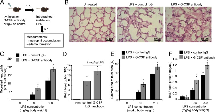
Blocking G-CSF activity aggravates LPS-induced acute lung inflammation. (A) Schematic representation of the experimental procedures. (B) Staining of lung sections shows emigrated neutrophils and polymerized fibrin in the pulmonary parenchyma. (C) The number of neutrophils emigrating to alveolar air spaces was quantified as volume fraction of the alveolar air space using standard point-counting morphometric techniques. (D) Neutrophil counts in BALF were calculated using the Wright–Giemsa staining method. (E) Pulmonary edema formation. (F) BALF total protein. The standard curve was constructed using BSA. Data shown are means ± SD of four experiments (n = 6 mice). *, P < 0.01 versus control (mice treated with IgG).

