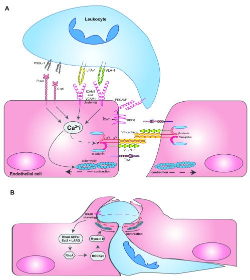
Figure 1. Opening and closing of endothelial junctions during diapedesis.
( A) Leukocytes interacting with several adhesion molecules on the endothelial cell surface trigger Ca 2+ signals inside endothelial cells, which are essential for leukocyte transmigration. It was reported that Ca 2+ signals triggered by the apical adhesion molecules were initiated by stores from the endoplasmic reticulum but that PECAM-1 Ca 2+ transients occurred rather local at transmigration sites through the TRPC6 channel. Ca 2+ signals trigger the activation of actomyosin-mediated pulling on endothelial junctions, influence the phosphorylation of components of the VE-cadherin-catenin complex, and trigger the recycling of the lateral border recycling compartment (LBRC) vesicle compartment. For a more detailed depiction of intracellular signaling steps, the reader is referred to recent reviews 7– 9. ( B) When leukocytes have already transmigrated more than halfway through the site of diapedesis, RhoA-mediated signaling triggered by the RHO guanine nucleotide exchange factors (GEFs) Ect2 and LARG stimulates ROCK2b, which activates actomyosin-based forces that support pore confinement, which leads to closure of the diapedesis pore 87. Abbreviations: ICAM1, intercellular adhesion molecule 1; LFA-1, lymphocyte function-associated antigen 1; PECAM-1, platelet and endothelial cell adhesion molecule 1; PSGL-1, P-selectin glycoprotein ligand-1; TRPC6, transient receptor potential canonical-6; VCAM1, vascular cell adhesion molecule 1; VE-cadherin, vascular endothelial cadherin; VE-PTP, vascular endothelial protein tyrosine phosphatase; VLA-4, very late antigen-4.

